
Anatomical Illustration: A Closer Look at the Lower Leg, Foot, and Achilles Tendon
Understanding the intricate structures of your lower leg, foot, and the vital Achilles tendon is key to appreciating how you move and how to care for these essential parts of your body.

The Foundation: Lower Leg Anatomy
Your lower leg is a powerhouse of movement, housing a complex interplay of muscles, bones, and connective tissues that allow for everything from a gentle stroll to a powerful sprint. This region is broadly divided into the anterior (front), lateral (side), and posterior (back) compartments, each with its own set of muscles responsible for specific actions.
Anterior Compartment Muscles
Dominating the front of your lower leg are the muscles responsible for dorsiflexion – lifting your foot upwards towards your shin. The most prominent here is the tibialis anterior. Imagine it as the primary driver for bringing your toes up when you walk or run, preventing your foot from scuffing the ground. Its tendon runs down the front of your ankle, often visible as a prominent cord, and inserts onto the medial (inner) side of your foot. This muscle group also plays a role in inverting your foot, turning the sole inwards.
Lateral Compartment Muscles
Situated on the outer side of your lower leg, the fibularis (or peroneus) muscles are crucial for everting your foot – turning the sole outwards – and stabilizing your ankle during activities like walking on uneven surfaces. The fibularis longus and brevis are the main players here. They originate from the fibula, the smaller bone in your lower leg, and their tendons wrap around the outside of the ankle bone before extending under the foot. This action is vital for preventing ankle sprains.
Posterior Compartment Muscles
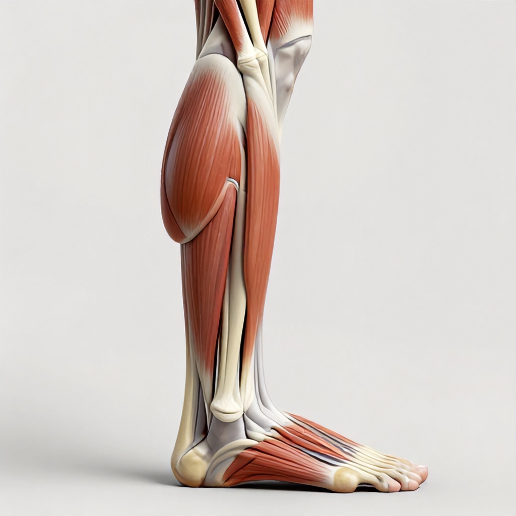
This is where the real power for pushing off the ground resides. The posterior compartment is further divided into superficial and deep layers. The superficial layer is dominated by the gastrocnemius and soleus muscles, which collectively form the calf. The gastrocnemius, with its two prominent heads, is the more superficial and visible muscle, giving the calf its shape. It crosses both the knee and ankle joints, contributing to plantarflexion (pointing your toes downwards) and also assists in flexing the knee. Beneath it lies the soleus, a broader, flatter muscle that also powerfully contributes to plantarflexion, but it only crosses the ankle joint. Together, these muscles are essential for walking, running, jumping, and standing on your tiptoes. The deep posterior muscles, like the tibialis posterior, flexor digitorum longus, and flexor hallucis longus, are deeper and control finer movements, including inverting the foot and flexing the toes.
Bone Structures of the Lower Leg
The skeletal framework of the lower leg consists of two long bones: the tibia and the fibula. The tibia, also known as the shinbone, is the larger and more medial of the two. It bears the majority of your body’s weight and articulates with the femur (thigh bone) at the knee and with the talus bone in the ankle. The fibula runs parallel to the tibia on the lateral side. While it doesn’t bear as much weight, it provides crucial attachment points for muscles and helps stabilize the ankle joint. The distal ends of both the tibia and fibula form the malleoli – the prominent bony bumps you feel on either side of your ankle, the medial malleolus (tibia) and the lateral malleolus (fibula).
The Intricate Foot Anatomy
Your foot is a marvel of biomechanics, a complex structure of 26 bones, numerous joints, ligaments, and muscles designed to support your body weight, absorb shock, and propel you forward. It’s divided into three main sections: the hindfoot, midfoot, and forefoot.
Hindfoot
This includes the talus and calcaneus bones. The talus is the keystone of the ankle, connecting the tibia and fibula to the foot. It articulates with the tibia and fibula above and the calcaneus below. The calcaneus, commonly known as the heel bone, is the largest bone in the foot. It bears a significant portion of your body’s weight during standing and walking and is the insertion point for the Achilles tendon. The articulation between the talus and calcaneus is crucial for inversion and eversion movements.
Midfoot
Comprising the navicular, cuboid, and three cuneiform bones, the midfoot forms the arch of your foot. These bones articulate with each other and with the hindfoot and forefoot, creating a flexible structure that acts as a shock absorber. The medial longitudinal arch, the most prominent arch, is particularly important for distributing weight and providing elasticity during movement.
Forefoot
This section includes the metatarsals and the phalanges (toe bones). There are five metatarsals, long bones that connect the midfoot to the toes. Each toe, except for the big toe (which has two phalanges), has three phalanges. The forefoot is critical for balance and propulsion, especially the big toe, which plays a significant role in the push-off phase of walking and running. The sesamoid bones, small pea-shaped bones embedded within tendons beneath the big toe joint, are also important for function and can be prone to injury.
Ligaments and Muscles of the Foot
A dense network of ligaments crisscrosses the foot, providing stability and holding the bones together. The plantar fascia, a thick band of connective tissue running along the bottom of the foot from the heel to the toes, is particularly important for supporting the arch and absorbing shock. Intrinsic muscles within the foot, though small, provide fine motor control and support for the arches. Extrinsic muscles originating in the lower leg also extend tendons into the foot to control its movements.
The Mighty Achilles Tendon
The Achilles tendon, also known as the calcaneal tendon, is the largest and strongest tendon in the human body. It’s a thick, cord-like structure formed by the merging of the gastrocnemius and soleus muscles in the posterior leg. This powerful tendon runs down the back of the ankle and inserts onto the posterior aspect of the calcaneus, the heel bone.
Function and Importance
The primary role of the Achilles tendon is to transmit the force generated by the calf muscles to the heel bone, enabling plantarflexion – the action of pointing your toes downwards. This movement is fundamental for walking, running, jumping, and even standing on your tiptoes. The tendon’s elasticity allows it to store and release energy, contributing to the efficiency of locomotion. Its strength is essential for absorbing the impact forces generated during high-impact activities.
Anatomical Detail and Clinical Relevance
Anatomical illustration of the Achilles tendon often depicts its fibrous structure and its direct connection to the gastrocnemius and soleus muscles. A semi-transparent overlay can be useful in medical illustration to show the tendon’s relationship to the underlying bone structures, particularly the calcaneus. The tendon itself has a relatively poor blood supply, especially in its mid-portion, which can make it slower to heal when injured. This vulnerability makes it susceptible to conditions like Achilles tendonitis, an inflammation of the tendon, and Achilles tendinopathy, a more chronic degeneration. Tears and ruptures, while less common, are also serious injuries that can significantly impact mobility. Understanding the tendon’s anatomical detail is vital for orthopedic and kinesiology professionals when diagnosing and treating these conditions. The directional flow of force through the tendon is also a key consideration in biomechanics and sports medicine.
Visualizing the Structures: Anatomical Illustration
Creating detailed anatomical illustrations of the lower leg, foot, and Achilles tendon requires a deep understanding of the musculoskeletal system. Medical illustrators use various techniques to render these complex structures accurately. A clinical aesthetic is often favored, emphasizing clarity and precision. This can involve using a clean color palette, clear line work, and precise shading to define the forms of muscles, bones, and tendons. For educational purposes, anatomical rendering might incorporate labels pointing to specific structures, highlighting origins and insertions of muscles, or showing the pathways of major nerves and blood vessels. A semi-transparent overlay can be a powerful tool, allowing the viewer to see deeper structures beneath superficial ones, such as visualizing the Achilles tendon’s attachment point on the calcaneus while still showing the overlying soft tissue. This level of anatomical detail is invaluable for students, healthcare professionals, and anyone interested in the mechanics of the human body. Whether for therapeutic purposes, understanding injury mechanisms, or simply appreciating the complexity of our anatomy, detailed anatomical illustration provides a clear and informative window into the lower extremity.