Clinical Facial Mole Removal and Professional Skin Safety
Clinical facial mole removal requires a professional medical assessment to distinguish between benign beauty marks and potentially harmful skin lesions. While many people seek removal for aesthetic reasons, the primary goal of a dermatologist is to ensure the health of the skin tissue before any cosmetic procedure begins. A facial mole, or nevus, is a cluster of pigmented cells that can vary in depth, size, and color. Because the face is a highly visible area with delicate skin, the methods used to remove these spots must prioritize minimal scarring and rapid healing. Professional intervention is the only way to guarantee that a suspicious growth is not a form of skin cancer, such as melanoma or basal cell carcinoma. Attempting to treat these spots at home leads to permanent damage.
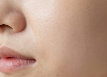
Understanding the Different Types of Facial Moles
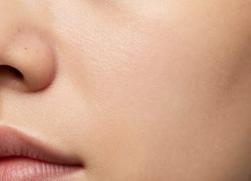
Not every spot on the face is the same. Some are flat and dark, while others are raised, flesh-colored, or even hairy. Doctors categorize these as junctional, compound, or intradermal nevi depending on where the pigment-producing cells sit within the skin layers. A junctional nevus is usually flat and brown. Compound nevi are slightly raised. Intradermal nevi are often skin-colored and sit higher on the surface. Identifying the specific type of skin lesion helps the dermatologist choose the most effective removal technique. It also determines how deep the procedure needs to go to prevent the mole from growing back. Most facial moles are benign, meaning they are non-cancerous, but any change in shape, size, or color warrants an immediate clinical evaluation.
The ABCDE Rule for Skin Health
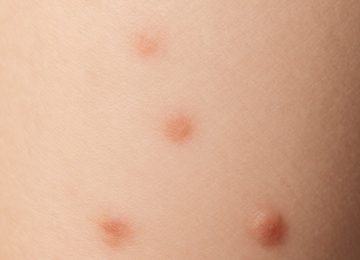
Dermatologists use a specific set of criteria to check for danger. Asymmetry is the first sign; one half of the mole does not match the other. Borders that are irregular, ragged, or blurred are also a red flag. Color variation, where the spot has shades of tan, brown, black, or even blue, requires attention. Diameter is another factor, as anything larger than a pencil eraser might be problematic. Evolving is perhaps the most important factor. If a mole changes over weeks or months, it must be biopsied. This systematic approach ensures that a cosmetic procedure does not accidentally overlook a serious health issue.
Effective Clinical Methods for Removal
Several safe and effective methods exist for removing facial moles in a clinical setting. The choice depends on the mole’s size, its depth, and its location on the face. A dermatologist will discuss the pros and cons of each approach to ensure the best aesthetic outcome.
Surgical Excision
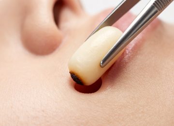
Surgical excision is a common choice for moles that sit deep within the skin layers. The doctor numbs the area with a local anesthetic and uses a scalpel to cut out the entire mole along with a small margin of healthy skin. This method is often necessary if there is any suspicion of malignancy because it allows the entire sample to be sent to a lab for a biopsy. After the mole is removed, the doctor uses very fine stitches to close the wound. On the face, these stitches are often placed with extreme precision to follow the natural tension lines of the skin. This helps the resulting scar blend in over time. It is a highly reliable way to ensure the mole is gone for good.
Shave Excision
For raised moles that do not appear suspicious, shave excision is a popular cosmetic procedure. The dermatologist uses a small, sharp blade to shave the mole off level with the surrounding skin. This technique is less invasive than a full excision and usually does not require stitches. The area is numbed first, so the patient feels nothing more than a bit of pressure. A small pink mark is left behind, which typically fades into a flat, white scar that is barely noticeable. Shave removal is excellent for “beauty marks” that catch on clothing or jewelry but do not have deep roots.
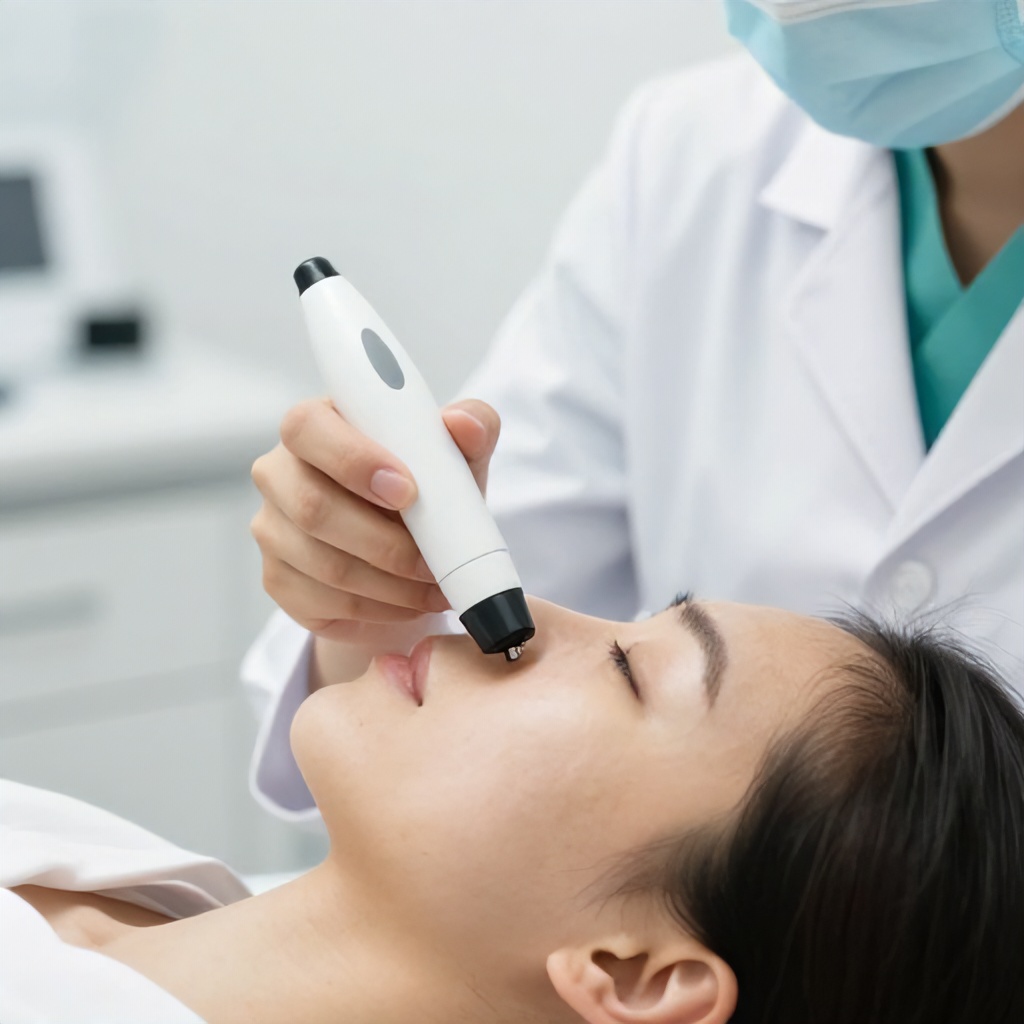
Laser Removal
Laser removal uses focused light energy to break down the pigment in the mole. This is best suited for small, flat, benign moles that are not deep in the dermis. The laser targets the dark melanin without damaging the surrounding tissue. It is a non-invasive option that carries a lower risk of scarring compared to cutting. However, laser treatment is not used for moles that need to be tested for cancer, as the light destroys the cells, making a biopsy impossible. Multiple sessions might be needed to completely clear the pigmentation from the skin spot.
Cryotherapy
Cryotherapy involves using liquid nitrogen to freeze the skin lesion. The extreme cold destroys the cells, causing the mole to eventually scab over and fall off. While very effective for skin tags and certain types of warts, it is used less frequently for deep facial moles because it is harder to control the depth of the freeze. There is a risk of leaving a white spot, or hypopigmentation, where the skin loses its natural color. Doctors usually reserve this for very superficial blemishes.
Electrocautery and Cauterization
Cauterization uses an electric current to burn away the mole tissue. This method seals the blood vessels as it works, which means there is very little bleeding. It is often used in combination with shave excision to smooth out the edges of the remaining skin. The heat from the tool kills the cells and prevents regrowth. Like cryotherapy, it is generally better for smaller, superficial spots rather than deep-rooted nevi.
The Importance of a Professional Biopsy
Every mole removed in a clinical setting should ideally be sent for a biopsy. This is a laboratory test where a pathologist examines the cells under a microscope to check for abnormalities. Even if a mole looks perfectly normal to the naked eye, microscopic analysis can reveal early signs of skin cancer. Skipping this step is a major risk. If a cancerous mole is partially removed or “burned off” without a biopsy, the remaining cancer cells can continue to grow beneath the surface, potentially spreading to other parts of the body. A dermatologist prioritizes your life over a quick cosmetic fix.
Why DIY Mole Removal is Dangerous
The internet is full of “natural” remedies like apple cider vinegar, garlic, or caustic mole removal creams. These are dangerous. These substances work by creating a chemical burn on the skin. They do not distinguish between the mole and the healthy skin around it, often leading to deep, permanent scarring and infections. Furthermore, if you attempt to remove a mole that is actually a melanoma, you are delaying life-saving medical treatment. You cannot see what is happening beneath the skin. A professional clinical setting provides a sterile environment, proper tools, and the expertise to handle complications. Saving a few dollars on a home kit is not worth a permanent facial scar or a missed cancer diagnosis.
Preparing for Your Appointment
Before heading to the dermatology clinic, take note of any changes you have seen in your skin. It helps to know how long the mole has been there and if it has ever bled, itched, or hurt. Avoid wearing heavy makeup to the appointment so the doctor can see the skin clearly. During the consultation, ask about the expected healing time and the likelihood of a scar. Most procedures are quick, often taking less than thirty minutes from start to finish. You will likely be able to drive yourself home and resume most normal activities immediately.
The Healing Process and Aftercare
Proper aftercare is the most significant factor in how well the skin heals after a facial blemish is removed. The goal is to keep the wound clean and moist. Dry wounds form thick scabs, which can pull at the edges of the skin and increase the size of the scar. Following the doctor’s instructions exactly will lead to the best aesthetic treatment results.
- Keep the area covered with a sterile bandage for the first 24 to 48 hours.
- Clean the wound gently with mild soap and water once or twice a day.
- Apply a thin layer of plain petroleum jelly or a prescribed antibiotic ointment to keep the site moist.
- Avoid picking at any scabs that form, as this introduces bacteria and damages the new skin growing underneath.
- Stay out of direct sunlight while the wound is fresh.
Managing Scarring on the Face
Scarring is a natural part of the body’s healing process, but there are ways to minimize its appearance on the face. Once the initial wound has closed and any stitches are removed, you can begin scar management. Silicone gel sheets or topical silicone gels are widely recommended by dermatologists. They create a protective barrier that hydrates the scar tissue and helps it flatten and fade faster. Massage can also help. Gently massaging the scar once it is fully healed breaks up collagen bundles that can make a scar feel hard or look raised. Patience is necessary. It can take twelve to eighteen months for a scar to fully mature and reach its final, faded appearance.
Sun Protection and Long-Term Skin Health
The new skin that grows after a mole removal is extremely sensitive to ultraviolet (UV) radiation. Sun exposure can cause the healing site to darken permanently, a condition known as post-inflammatory hyperpigmentation. Using a broad-spectrum sunscreen with at least SPF 30 is vital every single day, even when it is cloudy. Wear a wide-brimmed hat if you are going to be outdoors for an extended period. Protecting your skin from the sun doesn’t just help the scar; it prevents new moles and sunspots from forming. Skin health is a long-term commitment that goes far beyond a single procedure.
When to Call Your Dermatologist
While complications from clinical mole removal are rare, you should know what to look for during the healing process. If you notice increasing pain, significant swelling, or warmth around the site, these could be signs of an infection. Pus or a foul odor are also indicators that you need a follow-up. If the mole appears to be growing back, do not ignore it. Sometimes a few pigment cells are left behind, and they can regenerate. A quick check-up ensures that everything is progressing as it should.
Maintaining a Clear Complexion
Regular skin checks are the best way to maintain a clear and healthy complexion. Once a year, have a dermatologist perform a full-body skin exam. They have the tools, like a dermatoscope, to see deep into the layers of the skin and catch issues before they become serious. Between visits, perform your own skin checks at home. Use a mirror to look at your face, neck, and ears. If you find a new skin spot or a beauty mark that looks different than the others, get it checked. Taking a proactive approach to your skin care ensures that you stay healthy and feel confident in your appearance.
Facial mole removal is a straightforward process when handled by a professional. By choosing clinical methods over risky home treatments, you protect your skin’s integrity and your overall health. Modern dermatology offers various tools to ensure that the removal is as painless and scar-free as possible. Focus on the healing process, protect yourself from the sun, and listen to the guidance of your medical provider. Your face is your most visible feature, and it deserves the highest standard of care.