Spotting multiple skin growths on your foot sole can be concerning, and any new or changing skin abnormality in this area warrants a professional medical evaluation by a dermatologist or podiatrist. The skin on the plantar surface of your foot is unique, constantly subjected to pressure and friction, which can influence how various cutaneous lesions appear and behave. Understanding the different types of skin lesions that can develop here is crucial for your overall health education and early detection of potentially serious dermatological conditions.
Why Your Foot Sole Deserves Special Attention
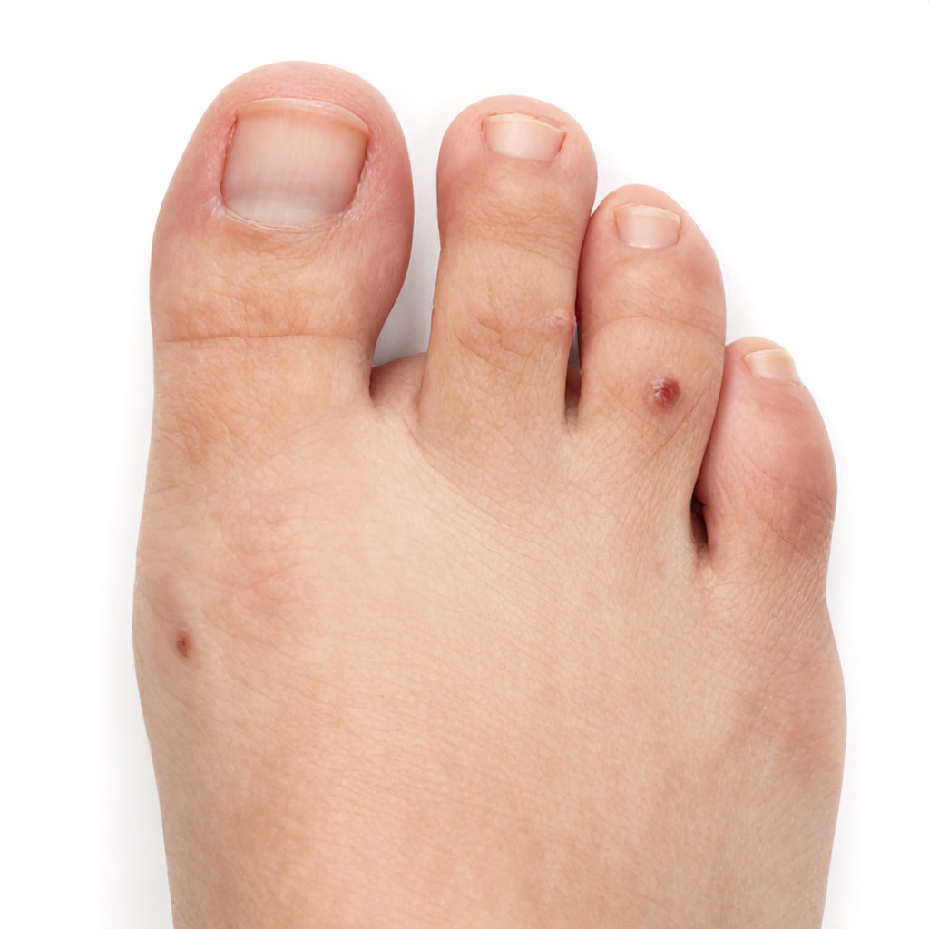
The skin on the bottom of your feet, known as the plantar surface, is distinct from skin elsewhere on your body. It’s thicker, designed to withstand significant pressure and impact, and it lacks hair follicles and sebaceous glands. This unique foot anatomy means that skin growths in this pedal region might look different or behave differently than those on other parts of your body. Because the foot sole is often out of sight, changes can go unnoticed for longer, making regular self-checks incredibly important.
The constant pressure from walking and standing can also alter the appearance of skin growths, sometimes making them flatter or causing them to grow inward. This can complicate self-diagnosis and even make clinical diagnosis more challenging without proper tools and expertise. That’s why professional assessment of any suspicious skin tumors or lesions is always recommended.
Common Benign Skin Growths on the Foot Sole
Many skin growths on the foot sole are harmless, or benign. While they might cause discomfort, they typically aren’t a threat to your health. However, even benign growths can sometimes mimic more serious conditions, so a definitive diagnosis is always best.
Plantar Warts (Verrucae)
Plantar warts are one of the most common types of epidermal lesions found on the foot sole. They are caused by the human papillomavirus (HPV) entering the skin through tiny cuts or breaks. Unlike warts on other body parts, plantar warts are often flattened by the pressure of walking and can grow inward, causing significant pain. They typically appear as grainy, flesh-colored or brownish bumps, sometimes with small black dots (which are tiny clotted blood vessels) at their center. Multiple warts can sometimes merge, forming a larger lesion called a mosaic wart. These verrucae can be quite stubborn and often require medical intervention for effective removal.
Corns and Calluses
These aren’t technically skin growths in the same way warts or moles are, but rather thickened areas of skin that develop in response to repeated pressure or friction. Calluses are broad, diffuse areas of thickened skin, usually painless, forming over bony prominences. Corns are smaller, more defined, and often cone-shaped, with a hard center that can press into the deeper layers of skin, causing sharp pain. They are common skin abnormalities, often caused by ill-fitting shoes or foot deformities. While not dangerous, they can be very uncomfortable and may require professional trimming or padding to relieve pressure.
Moles (Nevi)
Moles are common skin growths made up of pigment-producing cells called melanocytes. While most moles are benign, those on the foot sole, particularly on the plantar surface, warrant closer observation. Moles on the feet can sometimes be darker or more irregularly shaped than those on sun-exposed areas. It’s important to monitor any mole for changes, as even a seemingly innocent mole could potentially evolve into something more serious. A professional clinical view is always best for these particular skin lesions.
Seborrheic Keratoses
These are common, non-cancerous skin growths that often appear as we age. On the foot sole, they might be less typical in appearance than on other parts of the body. They usually look like waxy, “stuck-on” brown, black, or tan lesions with a slightly raised, rough surface. While generally harmless, if they become irritated, bleed, or change rapidly, they should be examined by a dermatologist to rule out other dermatological conditions.
Cysts and Lipomas
Cysts are sacs filled with fluid, air, or other material, and they can develop anywhere on the skin, including the foot sole. They often feel like small, movable lumps under the skin. Lipomas are benign fatty tumors that also present as soft, rubbery, movable lumps beneath the skin. Both are generally harmless but can cause discomfort if they grow large or are in a pressure-bearing area. Occasionally, they might require removal if they are painful or interfere with walking.
Potentially Malignant Skin Growths on the Foot Sole
While less common, malignant skin growths can occur on the foot sole. Because these areas are often overlooked, and because some types of skin cancer can present differently on the feet, early detection is absolutely vital. The good news is that when caught early, most skin cancers are highly treatable.
Melanoma
Melanoma is the most serious type of skin cancer, and it can occur on the foot sole, even in individuals with darker skin tones or those who haven’t had much sun exposure. A specific type, called acral lentiginous melanoma (ALM), frequently appears on the palms, soles, and under the nails. ALM often presents as a dark, expanding patch that may be mistaken for a bruise, mole, or fungal infection. It’s essential to be vigilant about any new or changing pigmented lesion on your foot. The classic “ABCDE” rule for melanoma still applies, though ALM can sometimes be amelanotic (lacking pigment), making it harder to spot. This serious health topic demands careful attention.
- Asymmetry: One half of the lesion does not match the other.
- Border irregularity: The edges are ragged, notched, or blurred.
- Color variation: The color is not uniform and may include shades of brown, black, tan, white, red, or blue.
- Diameter: The lesion is larger than 6 millimeters (about the size of a pencil eraser), though melanomas can be smaller.
- Evolving: Any change in size, shape, color, elevation, or any new symptom like bleeding, itching, or crusting.
Any suspicious pigmented lesion on the foot sole, especially one that meets any of the ABCDE criteria, requires immediate dermatological evaluation. Diagnostic imaging and biopsy are often necessary to confirm the diagnosis.
Squamous Cell Carcinoma (SCC)
Squamous cell carcinoma is another form of skin cancer that can develop on the foot sole, though it’s more commonly associated with sun-exposed areas. On the feet, SCC can sometimes arise from chronic wounds, scars, or areas of inflammation. It often appears as a firm, red nodule, a scaly patch, or a wart-like growth that may bleed or ulcerate. Unlike plantar warts, SCC tends to grow progressively and can be painful. It’s important not to dismiss a persistent, non-healing lesion as just a stubborn wart or callus.
Basal Cell Carcinoma (BCC)
Basal cell carcinoma is the most common type of skin cancer overall, but it is relatively rare on the foot sole. When it does occur, it might present as a pearly or waxy bump, a flat, flesh-colored or brown scar-like lesion, or a persistent sore that bleeds, oozes, or crusts and doesn’t heal. While BCC rarely spreads to other parts of the body, it can be locally destructive if left untreated. Its rarity on the pedal region means it might sometimes be misdiagnosed, highlighting the importance of expert clinical view.
When to Seek Professional Help for Foot Sole Growths
It’s always better to be safe than sorry when it comes to skin growths, especially on the foot sole. If you notice any of the following, schedule an appointment with a dermatologist or podiatrist promptly:
- A new growth that appears suddenly.
- Any existing growth that changes in size, shape, color, or texture.
- A growth that starts to bleed, itch, become painful, or develop an open sore.
- Multiple growths appearing without clear cause.
- A mole or lesion that looks asymmetrical, has irregular borders, varied color, or is larger than 6mm.
- A persistent lesion that doesn’t heal within a few weeks.
- Any growth that causes discomfort or interferes with walking.
Don’t try to self-diagnose or self-treat potentially serious skin abnormalities. A medical professional can provide accurate medical information and guide you on the best course of action.
What to Expect at the Doctor’s Office
When you visit a doctor for concerns about skin growths on your foot sole, they will typically perform a thorough examination. This might involve several steps to ensure an accurate diagnosis.
Physical Examination and History
Your doctor will visually inspect the affected area, noting the size, shape, color, and texture of the skin lesions. They will also ask about your medical history, including when you first noticed the growth, if it has changed, if it’s painful or itchy, and any family history of skin cancer. This initial clinical view helps them form an educated opinion.
Dermoscopy
Many dermatologists use a dermatoscope, a specialized handheld microscope, to examine skin growths in greater detail. This non-invasive tool allows them to see structures and patterns within the lesion that are not visible to the naked eye, providing valuable clues about whether a growth is benign or malignant. It’s a key part of modern dermatology practice.
Biopsy
If a growth looks suspicious, the doctor will likely recommend a biopsy. This involves removing a small sample of the tissue (or the entire growth, if small enough) and sending it to a pathology lab for microscopic examination. A biopsy is the definitive way to diagnose skin cancer and other complex dermatological conditions. There are different types of biopsies, including shave biopsies, punch biopsies, and excisional biopsies, chosen based on the nature of the lesion.
Diagnostic Imaging
In rare cases, especially if a growth is deep or suspected to be more invasive, diagnostic imaging such as ultrasound, MRI, or CT scans might be used to assess the extent of the lesion and whether it has affected deeper structures or spread. This is less common for typical epidermal lesions but can be important for larger skin tumors.
Prevention and General Foot Health
While you can’t prevent all skin growths, you can take steps to maintain good foot health and increase your chances of early detection.
- Regular Self-Exams: Make it a habit to check your feet, including the soles, between your toes, and under your nails, once a month. Use a mirror if needed to see all areas. Look for any new spots, changes in existing ones, or any sores that don’t heal.
- Protect Your Feet: Wear appropriate footwear that fits well to prevent corns, calluses, and friction blisters. When outdoors, consider applying sunscreen to your feet, especially if wearing open-toed shoes, as the pedal region can still get sun exposure.
- Maintain Good Hygiene: Keep your feet clean and dry to prevent fungal infections and other skin issues that could sometimes mask or complicate the appearance of other skin lesions.
- Be Aware of Your Family History: If you have a family history of melanoma or other skin cancers, you may be at higher risk and should be extra vigilant with self-exams and professional check-ups.
Taking a proactive approach to your foot health can make a significant difference in managing any skin abnormalities that may arise.
Understanding Your Diagnosis and Treatment
Once your doctor provides a diagnosis, they will discuss the appropriate treatment plan. For benign growths, treatment might involve observation, simple removal for comfort or cosmetic reasons, or addressing the underlying cause (like changing footwear for corns). For malignant growths, treatment options vary depending on the type and stage of cancer but often include surgical removal, cryotherapy, topical medications, or other specialized therapies. Early detection of malignant growths significantly improves treatment outcomes.
Remember, your feet carry you through life, and their health is an integral part of your overall well-being. Don’t hesitate to seek professional medical information and care for any concerns about skin growths on your foot sole.